Diagnostic Services
A consultation will be provided for patients referred for evaluation by their own medical ophthalmic or optometric physicians when questions arise regarding specific retinal contribution to a visual problem.
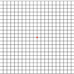
The Amsler Grid is used to monitor patients central visual field by using a grid of horizontal and vertical lines. Developed by Marc Amsler, the grid is a diagnostic tool that aids in the detection of visual disturbances caused by changes in the retina. To use the grid, the patient uses each eye separately a the small dot in the center. If the grid appears to have wavy lines or lines missing, the patient may suffer from macular disease.

Interocular pressure can be measured with Corneal Hysteresis machine (non-contact) Goodmann Applanation Tonometry, or Tono-pen. Elevated interocular pressure is a characteristic of glaucoma.
Evaluation of the visual fields can be performed using a variety of computerized program testing to assess both the extent and location of a defect in the visual field.
Computerized testing of color vision is an important measure of Optic nerve and Macular function. Screening of color vision can also be performed with Ishihara Color Test. Color testing is an important component of a comprehensive eye exam.
Evaluation of the anterior chamber angle may be required to evaluate various forms of glaucoma which can be associated with Retinal Disease.
The Slit Lamp allows for a binocular magnified view with focused illumination. This apparatus is widely used for a detailed examination of the front as well as the back of the eye. Special lenses may be required to focus on the back of the eye for Slit Lamp examination.
A binocular stereoscopic view of the inside of the eye can be obtained with a binocular indirect ophthalmoscope. Experience with this instrument allows the retina specialist to identify and assess a wide variety of disorders affecting the inside of the eye. Scleral Depression can be used in coordination with indirect ophthalmoscopy to visualize peripheral portions of the retina which would otherwise not be seen.
These evaluations allow a photographic assessment of retinal and optic nerve circulation. Computerization of the technology permits immediate evaluation of the findings. Both highly magnified and wide-angle camera can be utilized to examine the circulation of the retina.
Assessment of structural changes in the cornea are enhanced with computerized technology, which demonstrates a topographic evaluation of the cornea. These surface abnormalities can affect the visual acuity and quality.
This is an integral part of visual assessment. Computerized perimetry is utilized to measure peripheral blind spots and central visual field abnormalities. Results of this test may be diagnostic of retinal or optic nerve disorders as well as intracranial disorders including brain tumors or stroke.
This evaluation permits an assessment of electrical potentials related to functioning of the light sensitive components of the retina. It is useful both for diagnostic assessment and for ongoing follow-up of a variety of retinal disorders.
This non-invasive ultrasound evaluation can assess circulatory disorders involving the intracranial circulation. Many patients with vascular disorders involving the retina require further evaluation to rule out associated cerebrovascular abnormalities. Results of these tests are evaluated by an experienced radiologist.
Optical Coherence Tomography (OCT) permits measurement and assessment of retinal and optic nerve structure. Pre-and-subretinal pathologies can be assessed with this powerful computerized technology.
Measurement of farsightedness (hyperopia), nearsighted (myopia) and astigmatism (corneal asymmetry).
Measurement of the eyes’ need for glasses can be obtained using this instrumentation.