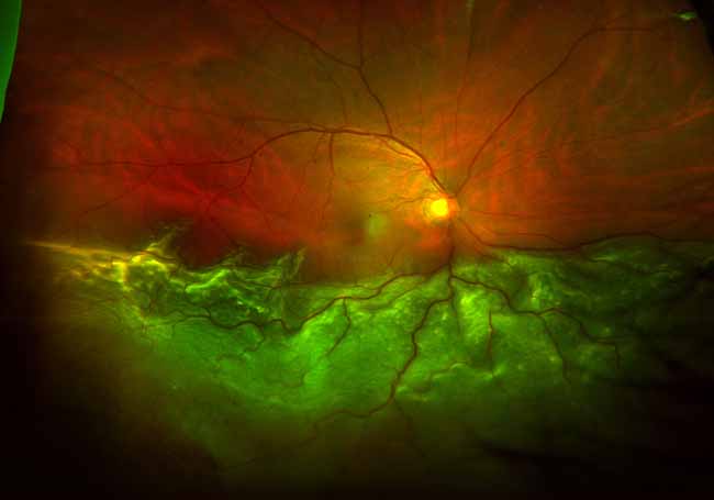
There are two major causes for retinal detachment: retinal tears and intraocular scar tissue. A tear in the retina may develop with or without previous injury and is frequently related to a variety of retinal degenerations.
A retinal examination using binocular indirect ophthalmoscopy, scleral depression and contact lens biomicroscopy permits early diagnosis of retinal degeneration, holes and tears which may be treated in our office facilities with laser or cryotherapy procedures to prevent development.
Certain retinal detachments may be treated in the office with pneumatic retinopexy which includes the intraocular injection of perfluorocarbon gas. Other cases require vitrectomy with or without scleral buckling. These procedures require operating room facilities. The surgeon may utilize intraoperative laser and heavy liquids to repair the detachment as well as intraocular gas or silicone oil.
Traction retinal detachments may be related to diabetic retinopathy, sickle cell retinopathy or chronic rhegmatogenous (tear related) detachment. These may require removal of intraocular scar tissue or areas of scarred retina (retinectomy). All Retina Group surgeons have completed both complete University residences in general ophthalmology as well as fellowship training in vitreo-retinal disorders. As their clinic practice is restricted to vitreo-retinal disorders, they have developed a unique expertise in the diagnosis and surgical treatment of these conditions.
Traction retinal detachments may be related to diabetic retinopathy, sickle cell retinopathy or chronic rhegmatogenous (tear related) detachment. These may require removal of intraocular scar tissue or areas of scarred retina (retinectomy).
All Retina Group surgeons have completed both complete University residences in general ophthalmology as well as fellowship training in vitreo-retinal disorders. Their continued specialization in vitreo-retinal disorders, allows them to develop a unique expertise in the diagnosis and surgical treatment of these conditions.


